
The Sleep Evidence, Part 2: Does Sleep Clear the Brain of Alzheimer's Proteins?
During deep sleep, the brain clears out metabolic waste, including the amyloid protein tied to Alzheimer's. It is one of the most compelling mechanisms in brain health, and one of the most overstated. Here is what the glymphatic system actually does, and what it cannot yet prove in people.
The essentials
The brain tissue itself has no lymphatic vessels, so it clears waste another way: fluid runs along its blood vessels and flushes the tissue.That flushing runs hardest during deep sleep, and it clears the very proteins tied to Alzheimer’s, amyloid and tau.A 2025 study identified the driver: slow waves of a brain chemical, norepinephrine, that make the arteries pulse and pump fluid through.A common sleeping pill, zolpidem, suppressed that pumping in mice even though it increased sleep time. Drugged sleep is not the same as natural sleep. (An animal finding, not a reason to stop a prescribed medication.)The human evidence is now real: deprive people of sleep and their amyloid and tau rise, and blunting deep sleep specifically raises amyloid the same night.It appears to matter most for people carrying the APOE4 gene, where poor sleep and genetic risk act on the same clearance machinery.Whether losing deep sleep predicts dementia in people is still unsettled: the sleep-stage statistics are mixed, even though the mechanism is real.The sturdier human signal is not a sleep stage but sleep continuity: unbroken sleep, and treated sleep apnea. That sets up the next post.
The brain cleans itself, mostly at night
Every other organ has lymphatic vessels, a drainage network that carries away cellular waste. The brain’s interior does not. So it improvised a different system, and the outline of how it works was only mapped in the last dozen years.
In a 2013 study1, researchers watching live mouse brains found something striking: when a mouse fell asleep, the tiny spaces between its brain cells opened up by about 60 percent. That widening let cerebrospinal fluid, the clear fluid around the brain, wash through the tissue far more freely than it could during waking. And what the fluid carried away included amyloid-beta, one of the proteins that builds up in Alzheimer’s disease. Clearance of amyloid ran faster in the sleeping brain than the waking one.
The plumbing behind this had been described a year earlier2. Fluid does not seep randomly through the brain; it travels along the outside of blood vessels, using them as channels to reach deep tissue, then drains back out along the veins. The system depends on tiny water channels, called AQP4, studded on the support cells that wrap the vessels. Remove those channels in mice and waste clearance drops by roughly 70 percent, and amyloid in particular stops being cleared properly. Because the network runs along the vasculature and behaves like the body’s lymphatic system, researchers named it the glymphatic system.
So the picture by the mid-2010s was: the brain flushes its own waste, the flushing runs on fluid moving along blood vessels, and it works far better asleep than awake. What nobody knew was why sleep flipped the switch.
What actually drives the flush
That answer arrived in 20253, and it is the new piece.
Using a new method to watch fluid move in mice that were sleeping naturally rather than anesthetized, the researchers found that during deep, non-dreaming sleep, a brain chemical called norepinephrine rises and falls in slow waves, roughly one cycle every fifty seconds. Norepinephrine tightens blood vessels, so each wave makes the arteries gently squeeze and release in the same rhythm. That slow, rhythmic pulsing of the vessels acts like a pump, driving the cerebrospinal fluid through the tissue. They confirmed it from both directions: stimulate the brain cells that release norepinephrine and the pumping strengthened; block it and the pumping stopped.
So the mechanism is not passive drainage. It is an active pump, and the thing running the pump is a brainstem rhythm that only organizes itself properly during natural deep sleep. That is why sleep, specifically, matters for clearance: the pump is a feature of the sleeping brain’s architecture.
The part about sleeping pills, handled carefully
The same study included a finding that is both striking and easy to misread, so it needs care.
The researchers gave mice zolpidem, the active ingredient in Ambien and one of the most common prescription sleep aids. The drug did what it is supposed to do: the mice fell asleep faster. But it suppressed the norepinephrine waves, and with them the pumping and the clearance. The sedative brought sleep on, but the flushing fell away with the waves. Drug-induced sleep, at least this kind, was not equivalent to natural sleep.
Two honest boundaries on this. First, it is a mouse study. The paper is explicit that we cannot yet measure this pump directly in people, so whether zolpidem does the same thing in humans is not established. Second, and this matters, this is not a reason to stop a prescribed sleep medication. It is a mechanistic finding about how natural sleep works, not clinical advice, and stopping a prescribed drug on the strength of a mouse study is exactly the wrong move. If the finding does anything useful, it is to reinforce that the goal is good natural sleep, and to give researchers a lead worth following. The place to discuss any sleep medication is with the clinician who prescribed it.
How much of this is actually true in people
A caveat has to sit alongside all of this, because the mechanism is compelling and the human evidence is thinner than it feels.
Almost everything above is animal work. The reason is technical: the imaging tools that let researchers watch fluid pump through a mouse brain cannot yet resolve the same flow in a human one. In people, we can see cerebrospinal fluid sloshing in the brain’s larger chambers during deep sleep, which is consistent with the mouse findings, but that is not the same as watching the deep-tissue flush itself.
What human researchers do instead is use indirect markers. The main one is a measurement taken from an MRI scan, called the ALPS index, that estimates how well fluid is moving along the spaces that surround the brain’s blood vessels. It is a surrogate, an inference about the pump rather than a picture of it, and the field is candid about that. But it lets researchers ask, in living people, whether worse glymphatic function tracks with Alzheimer’s pathology.
It does, and with a twist that ties back to genetics. In a 2025 study of older adults4, a lower ALPS index, meaning poorer estimated clearance, was linked to higher amyloid in the brain, but only in people carrying the APOE4 gene, the strongest common genetic risk factor for Alzheimer’s. In non-carriers the link was not significant. So the human evidence is real but indirect, and it suggests the clearance system matters most in exactly the people most genetically predisposed to accumulate amyloid.
That last point matters, and it comes back later. The theme across both mice and people is that APOE4 and the clearance system are entangled: the genetic risk does not act in isolation, it appears to act partly by making the brain’s overnight cleanup less effective.
The honest problem: deep sleep and dementia do not line up as neatly as the mechanism predicts
If the mechanism is right, the prediction seems obvious: people who get less deep sleep, the slow-wave stage when the pump runs hardest, should be at higher dementia risk. The human evidence on that specific prediction is mixed, and it is worth walking through honestly, because it is the place a careful reader would push back.
One study seemed to confirm it. In the Framingham Heart Study, researchers tracked how people’s slow-wave sleep changed over time5 and found that a faster decline in it was associated with higher dementia risk, roughly a 27 percent increase for each additional 1 percent lost per year. That fits the mechanism cleanly.
But the same cohort, in an earlier study6, pointed somewhere else. When those researchers looked at sleep stages at a single point in time, it was lower REM sleep, not slow-wave sleep, that predicted dementia, and non-REM stages showed no association at all. Same population, different measurement, different answer.
Then the largest test to date pooled five US cohorts, nearly 4,700 people, and found no consistent association between any sleep stage and dementia.7 If anything, in one subgroup, more slow-wave sleep was marginally associated with slightly higher risk, the opposite of the prediction, though the authors caution against reading much into it.
So how do these fit together? The most likely explanation, and the one the pooled study itself offers, is about what each measure captures. The physiology, the flush during deep sleep, is real and happens night to night. But a single overnight sleep study, measuring what percentage of one night someone spent in a given stage, is a crude and noisy snapshot. It may say more about last night than about a brain over decades.
The one finding that held up, the Framingham slow-wave result, measured change over many years rather than a single night, which may be why it caught a signal the snapshot studies missed. The mechanism and the population statistics have partly come apart, not because the mechanism is wrong, but because sleep-stage percentages are a blunt way to measure it.
There is a more useful signal buried in the same pooled analysis. Sleep stages did not predict dementia, but sleep disruption, fragmented, inefficient sleep with a lot of time awake in bed, and untreated sleep apnea did track with worse cognition. That points away from chasing a particular stage and toward the continuity of sleep, which is both more measurable and more actionable. It is also the thread that runs into the next post.
What happens when you actually deprive a human of sleep
The stage-percentage studies are noisy because they lean on a single night’s architecture. A cleaner test is experimental: take healthy people, disrupt their sleep on purpose, and measure the proteins directly. Three studies did versions of this, and together they are the most direct human evidence that the mechanism operates in people.
In the first, twenty healthy adults had their brains scanned for amyloid8 after a normal night and again after one night of total sleep deprivation. After the sleepless night, amyloid was measurably higher, in the hippocampus and thalamus, two regions hit early in Alzheimer’s. The increase was modest, around 5 percent, smaller than what is seen in established disease, but it was real and it appeared after a single night. Notably, this acute effect did not depend on whether the person carried the APOE4 gene, it happened across the board.
The second study got more specific about which part of sleep matters. Rather than keeping people fully awake, the researchers let them sleep but used quiet tones to selectively blunt their deep, slow-wave sleep9 without waking them. The morning after, the people whose slow-wave sleep had been most suppressed had the largest rise in amyloid in their spinal fluid, and the effect was specific to slow-wave sleep, not total sleep time. This is close to a direct human test of the mechanism: interfere with exactly the stage when the pump runs hardest, and amyloid goes up that same night.
Tau, the other Alzheimer’s protein, behaved differently, in a telling way. In that same study, tau did not budge from one disrupted night; instead, it was poorer sleep quality over the preceding several nights that tracked with higher tau. And in a separate line of work, one night of sleep deprivation raised spinal-fluid tau by more than 50 percent10, with the animal half of the study showing that sustained wakefulness also spreads tau pathology between connected brain regions. The pattern that emerges is that amyloid responds to a single bad night, while tau responds to the accumulation of many, which fits tau’s much slower turnover.
So the human experimental evidence is clearly supportive, and more convincing than the stage-percentage epidemiology, because it manipulates sleep directly and watches the proteins move. The honest boundary is that these are short-term studies of a protein marker, not proof that a lifetime of poor sleep causes dementia. They show the mechanism is live in humans. They do not, by themselves, close the loop to disease.
Why this may hit APOE4 carriers hardest
One thread has run quietly through all of this: APOE4, the strongest common genetic risk factor for Alzheimer’s, keeps turning up wherever clearance fails. The sharpest evidence for that connection comes from a 2023 mouse study.11 It was built to test what happens when the two biggest risk factors for late-onset Alzheimer’s, the APOE4 gene and disrupted sleep, are combined.
The researchers used mice carrying either the human APOE3 or the higher-risk APOE4 gene, and subjected them to chronic sleep loss. The effect depended entirely on the gene. Sleep deprivation increased amyloid buildup in the APOE4 mice but not in the APOE3 mice. The same sleep loss that did little on the lower-risk background drove pathology on the higher-risk one.
It worked through two failures, both matching the mechanism already described. Sleep loss disorganized the AQP4 water channels the clearance pump depends on, and it weakened the immune cells that normally wall off and contain amyloid plaques. Both happened only in the APOE4 animals.
This is mouse work, and it should be read as such. But it lines up with the human evidence from earlier, where poorer estimated clearance tracked with amyloid specifically in APOE4 carriers.
Two very different approaches, a precise mouse experiment and a human imaging study, point the same way. The clearance system and APOE4 are entangled, and sleep loss appears to matter most for the people already genetically prone to accumulate amyloid. For the roughly one in four people who carry a copy of APOE4, that is a reason to take sleep seriously rather than fatalistically. It is a direction the evidence points, not a proven rule.
A cycle that can feed itself
Put the pieces together and a loop appears. Wakefulness raises amyloid and tau and slows their clearance; over time, accumulating pathology damages the very brain regions that generate deep, consolidated sleep; worse sleep then clears even less. Each turn can reinforce the next. The same 2023 study saw a version of this directly: the mice with the most pathology went on to have the most disrupted recovery sleep.
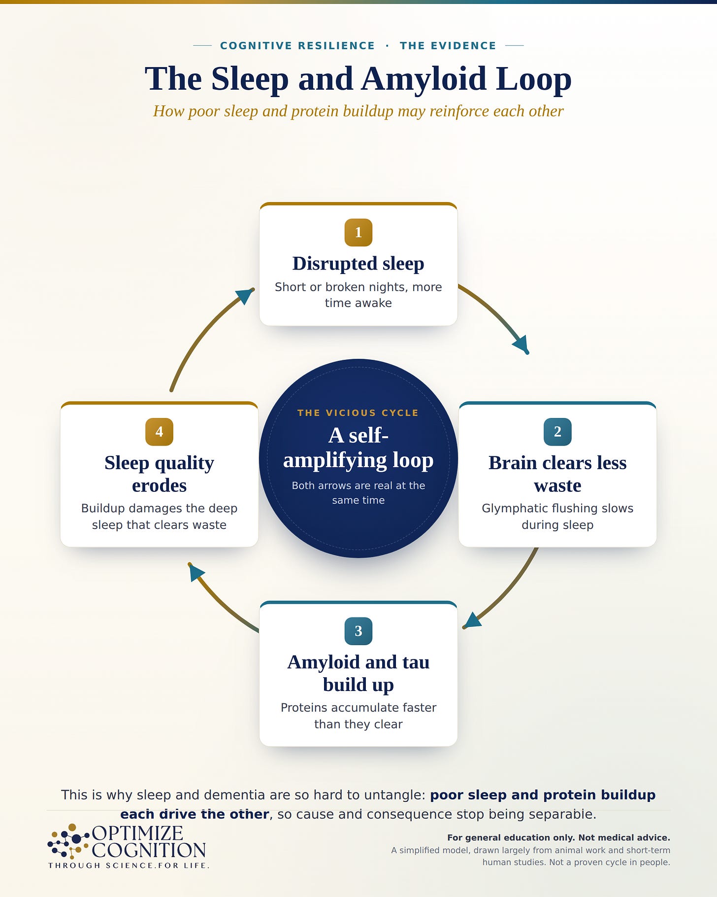
Two cautions keep this honest. A self-reinforcing loop does not mean an inevitable one, most people’s sleep varies for years without spiraling, and the loop is drawn largely from animal work and short human studies. And a loop cuts both ways: if poor sleep can feed the cycle, the reasonable hope, still unproven in a trial, is that protecting sleep helps interrupt it. That possibility, not a promise, is what makes the mechanism worth caring about.
Where this leaves things
The mechanism is one of the more satisfying stories in brain science: the sleeping brain runs a pump, driven by a slow chemical rhythm, that flushes out the very proteins that accumulate in Alzheimer’s, and when researchers disrupt it, those proteins rise. Worked out first in mice, it now has real human support, deprive people of sleep and their amyloid and tau move in the predicted direction, and it appears to matter most in those genetically most at risk.
But the honest reader should hold two things at once. The acute physiology is real and increasingly well demonstrated. The leap from that physiology to “poor sleep over a lifetime causes dementia” is not proven, and the human epidemiology on sleep stages is mixed, which is why this post spent as long on what does not line up as on what does. The mechanism tells you why sleep could matter for the brain. It does not, on its own, tell you how much protecting your sleep will change your future, and no trial has yet answered that.
What the mechanism does justify is taking sleep seriously as more than rest, and it points to the part that seems to matter most: not chasing a particular sleep stage, but protecting continuous, unbroken sleep and treating the disorders that fracture it. That is where the next post goes, to the sleep disorders that carry the clearest signal, apnea chief among them, and what the evidence says about doing something about them.
References
- Xie L, Kang H, Xu Q, et al. Sleep drives metabolite clearance from the adult brain. Science. 2013;342(6156):373-377. https://doi.org/10.1126/science.1241224
- Iliff JJ, Wang M, Liao Y, et al. A paravascular pathway facilitates CSF flow through the brain parenchyma and the clearance of interstitial solutes, including amyloid β. Sci Transl Med. 2012;4(147):147ra111. https://doi.org/10.1126/scitranslmed.3003748
- Hauglund NL, Andersen M, Tokarska K, et al. Norepinephrine-driven slow vasomotion drives glymphatic clearance in sleep. Cell. 2025;188(3):606-622.e17. https://doi.org/10.1016/j.cell.2024.11.027
- Kang KM, Park C, Byun MS, et al. APOE4 modulates the association between DTI-ALPS index and Alzheimer’s pathologies. Alzheimers Dement. 2025. https://doi.org/10.1002/alz.70837
- Himali JJ, Baril AA, Cavuoto MG, et al. Association between slow-wave sleep loss and incident dementia. JAMA Neurol. 2023;80(12):1326-1333. https://doi.org/10.1001/jamaneurol.2023.3889
- Pase MP, Himali JJ, Grima NA, et al. Sleep architecture and the risk of incident dementia in the community. Neurology. 2017;89(12):1244-1250. https://doi.org/10.1212/WNL.0000000000004373
- Yiallourou SR, Cavuoto MG, Bei B, et al. Sleep architecture and incident dementia in the Sleep and Dementia Consortium. Sleep. 2025;48(9):zsaf146. (Pooled 5-cohort analysis, n=4,657.)
- Shokri-Kojori E, Wang GJ, Wiers CE, et al. β-Amyloid accumulation in the human brain after one night of sleep deprivation. Proc Natl Acad Sci USA. 2018;115(17):4483-4488. https://doi.org/10.1073/pnas.1721694115
- Ju YS, Ooms SJ, Sutphen C, et al. Slow wave sleep disruption increases cerebrospinal fluid amyloid-β levels. Brain. 2017;140(8):2104-2111. https://doi.org/10.1093/brain/awx148
- Holth JK, Fritschi SK, Wang C, et al. The sleep-wake cycle regulates brain interstitial fluid tau in mice and CSF tau in humans. Science. 2019;363(6429):880-884. https://doi.org/10.1126/science.aav2546
- Wang C, Nambiar A, Strickland MR, et al. APOE-ε4 synergizes with sleep disruption to accelerate Aβ deposition and Aβ-associated tau seeding and spreading. J Clin Invest. 2023;133(14):e169131. https://doi.org/10.1172/JCI169131
This article is for general education, not medical advice, and reading it does not create a physician-patient relationship. It reports research findings, most of them from animal studies or short-term human experiments, which show mechanisms and associations rather than proof that sleep changes a person's dementia risk. Before changing anything about your sleep or medications, talk with your own clinician, who can weigh what applies to you.